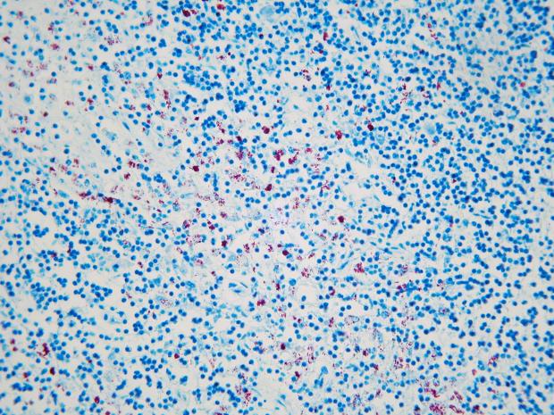
A case of Johne’s disease (Mycobacterium avium subsp. paratuberculosis (MAP)) was recently diagnosed in a 7 month old Friesian heifer submitted for postmortem examination to AFBI Stormont.

The calf was submitted for postmortem after a history off ill thrift, weight loss and death. According to the history provided there were also several other calves on the farm with similar clinical signs.
At postmortem examination the calf had a low Body Condition Score (BCS) of approx. 1.5 and weighed 104kg. There was faecal staining of the perineum, and there was scant small intestinal contents with watery brown contents in the caecum and large intestine. Other findings included some ulceration of the tongue and some cranioventral lung consolidation affecting approximately 10% of both lungs.
Further samples were taken at postmortem for diagnostic testing including for bacteriology, parasitology, virology and histology.
Bacteriology findings were negligible. Faecal egg counts were negative for liver fluke, nematodirus, rumen fluke, stomach worms and coccidia. Lungworm larvae however were detected in faeces. Respiratory viral testing for PI3V, BRSV and IBR were negative as was testing for BVD. Histology of the lung under the microscope showed a mild bronchopneumonia, while histological evaluation of iliocaecal junction revealed a chronic granulomatous enteritis. Specialised histological Ziehl-Neelsen staining of the iliocaecal junction and lymph node showed extensive acid fast bacteria present consistent with Johne’s Mycobacterium avium subsp paratuberculosis (MAP) infection. A faecal PCR test was performed for MAP and was positive. This animal demonstrated clinical signs of Johne’s and was positive on histological examination and faecal PCR.

Signs of Johne’s disease are typically seen in animals that are between 3 and 5 years old but can occasionally be seen in animals that are younger than two years of age. As the animal gets older, the signs become more obvious. An infected animal may also have increased susceptibility to other disease before the obvious signs occur.
There is no effective treatment or vaccination for Johne’s disease.
The direct economic impact on farms will vary between farms and depends upon the proportion of cattle infected and the extent to which infection has progressed in infected cattle. Infection can contribute to reduced milk yield and increased susceptibility to other conditions such as infertility even before the more obvious signs of disease appear. Infected cattle may be culled due to poor productivity before a diagnosis of Johne’s disease. Therefore, Johne’s disease can be an unrecognised cause of excessively high cull rates.
AFBI’s CHECS Cattle Health Scheme provides a pathway to manage and control Johnes Disease at farm level, and in Northern Ireland there is also a voluntary, industry led control programme managed by Animal Health and Welfare NI.
Due to the age of this calf Johne’s disease was not high on the list of differential diagnoses and only through submission for postmortem to AFBI was the disease confirmed, demonstrating that Johne’s disease must be kept in mind as a differential for calves of this age especially if a known history of Johne’s on farm.
This case demonstrates the value of submitting animals for postmortem. The diagnosis of disease and surveillance for other potential diseases, not only has value at farm level but also added value for the wider livestock sector.
For further information on Johne’s disease, the importance of control, diagnosis of Johne’s and the principles of control, contact your local veterinary practitioner, AFBI Cattle Health Scheme and Animal Health and Welfare NI.
Notes to editors:
AFBI’s Vision is “Scientific excellence delivering impactful and sustainable outcomes for society, economy and the natural environment”.
AFBI’s Purpose is “To deliver trusted, independent research, statutory and surveillance science and expert advice that addresses local and global challenges, informs government policy and industry decision making, and underpins a sustainable agri-food industry and the natural and marine environments”.
AFBI’s core areas:
- Leading improvements in the agri-food industry to enhance its sustainability.
- Protecting animal, plant and human health.
- Enhancing the natural and marine environment.
Latest news
- Soil Health Research to be Featured at AFBI Open Days in June 15 May 2024
- AFBI Open days - Improving the environmental sustainability of dairy farming through improved nutrition 14 May 2024
- AFBI Open days – How to make the most of early life to improve your future dairy herd 07 May 2024
- Public Appointment Vacancy - Chairperson to the Board of the Agri-Food and Biosciences Institute (AFBI) 02 May 2024
